SASS Clinical Note #5: Fibrovascular Polyp
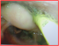
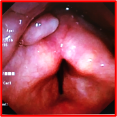
The picture on the left was sent to SASS by BH & LF who discovered this mass while administering a FEES study. The picture on the right was sent to SASS long ago by an SLP. Polyps are relatively rare in the pharynx but not the larynx. More often cysts are found in the pharynx. With both masses, their surfaces are smooth, and often whitish in color unlike malignant tumors that are irregular in shape and have rough, often angry, surfaces. Polyps tend to be softer and more fluid-like, while cysts are more fixed and dome-shaped. The tumor shown above has been confirmed by Otolaryngology to be a fibrovascular polyp (FVP). Close examination of its surface will confirm a dense vascular supply. These masses develop in the upper digestive tract, are benign and slow growing, usually pedunculated, and relatively rare. Their etiology is unknown but more often originate as small mucosal tumors adjacent to the cricopharyngeal muscle. Those found higher up in the hypopharyngeal cervical area are very rare. If large enough, they may be regurgitated into the pharynx or even the oral cavity, but it is difficult to determine its growth origin location without direct laryngoscopy investigation. The medical literature is sparse, but it appears to occur more in males than females (9:4) in their 60’s and 70’s. Tare reported cases in infants.
Most patients are completely asymptomatic leading to surprise discoveries during FEES. If the FVP is large enough or in a position to block or restrict passage of bolus material into the cervical esophagus, obstructive dysphagia may result. Some reports have described these polypoid masses to be so large as to cause dyspnea, and sudden asphyxia from laryngeal blocking. The latter issue is the most concerning to ENTs. Surgical removal is the primary treatment, and recurrence or malignant transformation are rare.
Gupta, M., Chaudhary, N., & Gupta, M. (2011). Fibrovascular polyp of the
oropharynx. Singapore medical journal, 52(2), e35–e36.
Haytoglu, S., Tuhanioglu, B., Bozkurttan, A., & Arikan, O. K. (2015). Giant Hypopharyngeal Fibrovascular Polyp: A Case Report and Review of the Relevant Literature. Case reports in otolaryngology, 2015, 670302. https://doi.org/10.1155/2015/670302
I, H., Kim, J. S., & Shim, Y. M. (2006). Giant fibrovascular polyp of the hypopharynx: surgical treatment with the biappoach. Journal of Korean medical science, 21(4), 749–751. https://doi.org/10.3346/jkms.2006.21.4.749
Oka, M., Ueha, R., Nito, T., & Yamasoba, T. (2016). Giant fibrovascular polyp in the hypopharynx: a case report and review of the literature. SpringerPlus, 5(1), 1443. https://doi.org/10.1186/s40064-016-3144-y